XERF is coming to Dermique! Get early access with exclusive savings of up to 20%. Limited spots available. See the offer →

Moles and dermoscopy

Moles are common skin growths, but some require closer monitoring. Changes in a mole’s size, shape, color, or appearance should never be ignored.

At Dermique, we perform dermoscopic evaluations of moles and other skin lesions to detect suspicious changes at an early stage. Our approach is medical and precise — we first examine the lesion and then determine whether it should be monitored, removed, or evaluated further.

See more…

Types of moles we evaluate

Red moles and angiomas

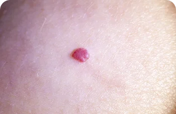
So-called “red moles” are most commonly cherry angiomas — benign vascular growths that may develop with age or due to genetic predisposition. They are generally harmless but should be evaluated if they grow, bleed, are frequently irritated or injured, or become an aesthetic concern.

Following evaluation, we may recommend monitoring or removal with DermaV by Lutronic.

Red moles and angiomas

Red moles and angiomas – small red spot on the skin

So-called “red moles” are most commonly cherry angiomas — benign vascular growths that may develop with age or due to genetic predisposition. They are generally harmless but should be evaluated if they grow, bleed, are frequently irritated or injured, or become an aesthetic concern.

Following evaluation, we may recommend monitoring or removal with DermaV by Lutronic.

Common melanocytic moles

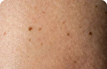
Melanocytic moles are the most common type of mole found on the face and body. Most are benign, but they should be monitored for changes such as growth, changes in color, asymmetry, bleeding, or itching.

Common melanocytic moles

Common melanocytic moles – skin with small brown moles

Melanocytic moles are the most common type of mole found on the face and body. Most are benign, but they should be monitored for changes such as growth, changes in color, asymmetry, bleeding, or itching.

Types of moles we evaluate
Mole removal

Mole removal is performed only after a dermoscopic evaluation. Depending on the type, size, and location of the mole, the dermatologist will determine the most appropriate removal method to achieve an optimal aesthetic result.

Dermoscopy

Dermoscopy is a non-invasive diagnostic technique that allows for a detailed examination of moles and other skin lesions. Using a specialized magnifying device called a dermatoscope, a dermatologist can evaluate structures and patterns beneath the skin’s surface that cannot be adequately assessed with the naked eye.

Dermoscopy can help identify suspicious changes at an early stage and allows for a more accurate assessment of whether a mole should be monitored, removed, or sent for histopathologic examination.

A routine dermoscopic skin examination is generally recommended once a year. Patients with numerous moles, fair skin, a family history of melanoma, a history of atypical moles, or significant sun exposure may require more frequent examinations based on their individual risk.

Start with a comprehensive skin evaluation and a personalized treatment plan from Dermique’s medical team.
Frequently asked questions
When should I be concerned about a mole?
Use the ABCDE rule when checking your moles:

A — Asymmetry: one half does not match the other

B — Border: irregular, uneven, or poorly defined borders

C — Color: uneven or multiple colors within the same mole

D — Diameter: larger than 6 mm

E — Evolving: any change in size, shape, color, or appearance

If you notice any of these signs, see a dermatologist promptly for evaluation.
For patients without additional risk factors, a dermoscopic examination is recommended once a year. Patients with numerous moles, fair skin, or a family history of melanoma should be evaluated every six months.
Mole removal is performed under local anesthesia and is completely painless. Recovery is quick and, in most cases, does not require any downtime.
Not every mole needs to be removed, and not every mole is suitable for the same removal method. The decision is made after a dermoscopic evaluation by a dermatologist, taking both medical and aesthetic considerations into account.

Atrophic scars

Atrophic scars are indented scars that most commonly appear following acne or other inflammatory conditions. They are caused by a loss of collagen and uneven skin regeneration.

Appropriate treatment methods

At Dermique, we don’t treat acne scars using just one technique. Where necessary, we combine: